SPH405
| Neurological Foundations of Speech, Language and Hearing |
![]()
![]()
![]()
![]()
![]()
![]()
Neurological Basis for Somesthesis
GOAL: TO DEVELOP GENERAL APPRECIATION OF THE NEUROLOGICAL BASIS
FOR SOMESTHESIS.
Apply knowledge of A/P to the evaluation of the peripheral speech mechanism.
Differentiate the types of somesthetic senses
Describe the three neuron organization of the somesthetic system.
There are three types of Somesthesis : mechanoreception; thermoreception; nocireception.
Mechanoreception is the sense of gross position changes body tissues.
- Neurons are mechanically displaced by some external force.
- Mechanoreception includes several kinds of displacement
- The TACTILE ("touch") sensations of touch, pressure or vibration.
- The KINESTHETIC or PROPRIOCEPTIVE sense of position and movement.
Thermoreception is the sensation of heat or cold.
Nocireception is pain associated with tissue destruction.
There are gradations of the somesthetic sensory experience.
- Not all sensations fit into the groups of "pain, temperature, touch."
- For example: into which of the above categories would you place: itch or tickle, or stereognosis?
- Perhaps each individual has special percepitions of these stimuli.
- The somesthetic experience is graded because stimuli impinge on receptors in groups.
- Stimulation of a single receptor is rare.
- Reality of touch is quite complex, and temporal and spatial summation result in great variations in the type of sensation.
Special somesthetic receptors fall into two braod categories: exterioceptors and enteroceptors.
Exterioceptors are located in skin, joints, muscles and tendons
- CUTANEOUS RECEPTORS are located just beneath the skin.
- They sense exterioceptive changes: changes in the external surface of the body.
- These sensations include touch pressure, pain, vibration, and thermal sensitivity.
- PROPRIOCEPTORS are located in the striated muscles, ligaments and joints.
- They provide continuous feedback on muscle stretch status.
- This feedback in extremely important because it enables modification of movement in progress.
Exteroceptors are linked to the CNS by SOMATIC AFFERENT
NEURONS.
- Not all of the input is conscious
- Responses to the unconscious input may be reflexive.
ENTEROCEPTORS respond to changes in the internal viscera and blood
vessels.
- They are associated with autonomic NS.
- Include vasosensory receptors.
- Vasosensory receptors respond to tension between oxygen and carbon dioxide levels in the blood stream.
- Their input to the afferent ANS gives initiates the ventilation response.
Somesthetic receptors can be categorized according to their sensitivity and adaptability.
Sensitive receptors are readily excited.
- They readily adapt and then their response drops off.
- The most sensitive and adaptive of the receptors are ENCAPSULATED.
- Encapsulated receptors have concentric layers of tissue around the nerve ending.
- They generate an action potential when capsule is deformed.
- Encapsulated receptors are located in the lips. fingers as well as other body parts. (Examples of encapsulated receptors are Meissner Corpuscles & Pacinian Corpuscles).
- Moderately Sensitive are "Expanded Tip Receptors." These receptors convey sensation from the dermatomes and the joiints. They are moderatey adapting.
- The least adaptive receptors are free of surrounding capsules and not as sensitive. Accordingly, they are called "Free" receptors.
- Free receptors are arborized throughout skin and viscera.
- They sense gross temperature variations and pain.
Cutaneous somesthesis is segmented in DERMATOMES.
- Dermatomes are topical regions of touch sensitivity.
- They correspond to spinal or cranial nerve segments.
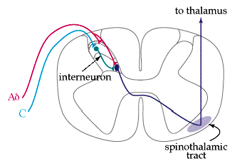
All somesthesis is transmitted to the CNS via the THREE NEURON arrangement.
FIRST ORDER neurons begin at the receptors.
- Receptors have their cell bodies in the dorsal root ganglia of the cord.
- They synapse in posterior horn gray of the spinal cord or in the lower levels of the brainstem.
SECOND ORDER neurons decussate (cross to the other side of the
CNS).
- Through the anterior white commissure of the cord or through the lower part of the brainstem.
- They turn rostrally...
- and ascend to thalamus.
THIRD ORDER neurons in ventral or posterior nuclear group to radiate to
somesthetic cortex.
- Primary reception is in the postcentral gyrus.
- Association occurs in the paracentral gyrus...
- ...of the parietal lobe.
FIRST ORDER neurons begin at the receptors.
- Receptors have their cell bodies in the dorsal root ganglia of the cord.
- They synapse in posterior horn gray of the spinal cord .
SECOND ORDER neurons have cell bodies in the posterior horn gray.
- Their axons decussate through the anterior white commissure of the cord and cross to the anterior half of lateral column.
- They then turn rostrally.
- They ascend to thalamus. In the brainstem and thalamus, the axons aggregate with the axons from the posterior column-lemniscal system to form the Medial Lemniscus.
THIRD ORDER neurons cell bodies lie in the ventral nuclear group of the
thalamus and radiate to somesthetic cortex.
- They terminate in the postcentral gyrus
- With association fibers in the paracentral gyrus
- Of the parietal lobe.
Lesions affecting the second or third order neurons of the spinothalamic tract will produce unilateral paresthesia on the contralateral side.
TRIGEMINOTHALAMIC tracts convey somesthetic impulses from the facial dermatomes to the brain.
These somesthetic impulses include fine touch as well as gross touch, but the routes of the two types of sonesthesis are not the same.
GROSS TOUCH FROM FACE:
FIRST ORDER NEURONS have cell bodies in the semilunar
ganglion, and axons course from the periphery to the mid-pons
level of the brainstem.
These axons form the descending tract of V.
- This tract travels caudally through the lower lateral pons and lateral medulla to C-1 thru C-4.
- They synapse at Nucleus of Descending Tract of V ain segments C-1 through C-4.
SECOND ORDER NEURONS have cell bodies in the nucleus of
the descending tract of V and send axons across the midline.
These axons then turn and travel rostrally...
- Forming the ventral ascending tract of V.
- They pass through lateral medulla, pons and midbrain forming the trigeminal lemniscus.
- Axons end in the thalamus
THIRD ORDER NEURONS have their sell bodies in the ventral
nuclear group of the thalamus. lateral course.
The axons take a lateral course from the thalamus to the...
- Posterior limb of internal capsule.
- They terminate in the postcentral gyrus of theparietal lobe.
FINE TOUCH FROM FACE:
FIRST ORDER NEURONS have their distal processes in
distribution of V to the facial dermatomes.
- The cell bodies are in the semilunar ganglion.
- Axons synapse in Chief Sensory Nucleus of V at the mid pons level.
SECOND ORDER NEURONS have cell bodies in Chief Sensory
Nucleus of V.
- They send axons rostrally in Dorsal Secondary Ascending Tracts of V.
- Some of these axons cross, some do not.
- They all synapse in the posterior portion of ventral nuclear group of thalamus.
THIRD ORDER NEURONS have their cell bodies in the ventral
nuclear group of the thalamus.
- They send axons to the posterior limb of the internal capsule.
- These axons terminate in the facial area of somesthetic cortex.
Unilateral lesions affecting the second and third order neurons of the trigeminothalamic tracts may produce contralateral gross touch symptoms, but the bilateral representation of the fine touch tracts may mask these symptoms and make them clinically undetectable.
The POSTERIOR COLUMN LEMNISCAL SYSTEM conveys fine touch impulses from the periphery of the body, especially the extremities, to the brain.
FIRST ORDER neurons at the periphery have specialized nerve endings
to be sensitive to fine touch. Their cell bodies are located in the in
posterior root ganglia of the cord.
- AXons course into the posterior columns of cord and turn in a rostral direction, traveling up the cord all the way to the brainstem..
- At caudal medulla, they synapse at nucleus gracilis (lower ext) and nucleus cuneatus (upper extremity).
SECOND ORDER neurons have their cell bodies in the Nuclei Cuneatus and
Gracilis near the Spinobulbar Junction.
- Their axons cross midline to form the "Sensory Decussation."
- The collective second order axons join the spinothalamic tracts to form the Medial Lemniscus.
- This bundle of axons courses through brainstem and into the thalamus.
THIRD ORDER neurons have their cell bodies in ventral nuclear group of
dorsal thalamus.
- Axons pass into the posterior limb of the internal capsule.
- They terminate in the postcentral Gyrus.(Somesthetic Cortex of Parietal Lobe)
Once you have finished you should:
Go on to Web
Activity 1
or
Go back to Somatosensory System
E-mail Bill Culbertson
at bill.culbertson@nau.edu
Call Bill Culbertson
at (520) 523-7440
Copyright © 1999
Northern Arizona University
ALL RIGHTS RESERVED