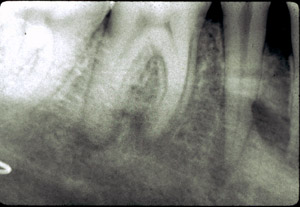
History: A 42 year-old white male had an intraoral full mouth x-ray examination by a local dentist prior to construction of a partial denture. The patient had no oral complaints other than being partially edentulous. The x-ray examination revealed bilateral impacted third molars, each associated with a cystic defect which extended beyond the limits of the intraoral film.
Clinical: Clinical examination showed a partially edentulous mandible with no expansion of the mandibular anatomy. There was no pain and only firm bone could be palpated. X-ray examination revealed bilateral impacted lower third molars each associated with a cystic lesion; the right side was 3 cm in diameter; the left side extended up into the ramus of the mandible and the impacted molar was located near the angle of the mandible.