Lesion Description Practice
Here are several lesion with descriptions to assist you in writing a description.
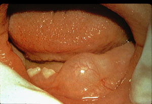
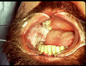
1. A large pink, round, fluid filled lesion on the gingival
adjacent to # 24, approximately 20 x 20 mm. Patient reports it appeared when
the tooth under it was coming through the gums. It is tender.
Color
Consistency
Size
Texture
Appearance
Location
History/Symptoms
2. A 7x5 mm oval, unilocular, radiolucency with well defined
borders between # 23 and 24.

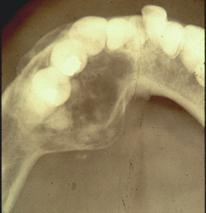
3. A larger radiolucency with well defined borders, fill
with multiple radiopacities in the area # 20-24 causing expansion of the bone.
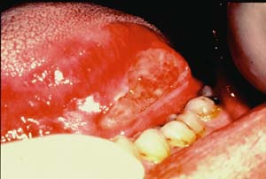
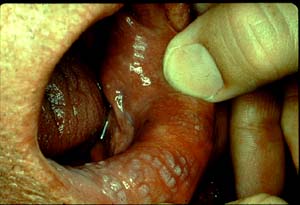
4. A large red and white, irregular shaped, ulcerated lesion
on the left lateral border of the tongue, approximately 25x10 mm.
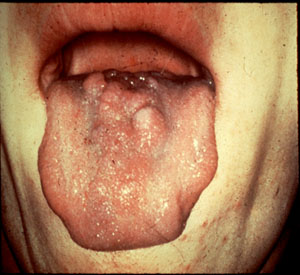
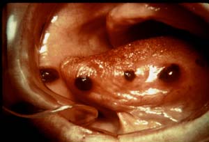
5. Six, pink, firm nodules on the dorsal surface of the
tongue adjacent to the cirumvalate papilla, each is approximately 6x6 mm in
size.
6.A large white plaque, with a rough texture that does not
wipe off covering the entire right buccal mucosa.
7. A large, slightly raised, white corrugated lesion covering
the labial mucosa in the area of # 26-27.
8. 4 lesion: 1 a soft, smooth, redish-brown, oval 2x3x1 lesion
on rt buccal mucousa: 2. a soft, smooth, redish-brown, oval 2x3x1 lesion on
lft anterior lateral boarder of the tongue: 3. a soft, smooth, redish-brown,
oval 1x1x1 lesion on lft anterior lateral boarder of the tongue slightly posterior
to #2: 4. a soft, smooth, redish-brown, oval 3x4x1 lesion on lft lateral boarder
of the tongue slightly posterior to #3. All lesions appear to be blood filled.
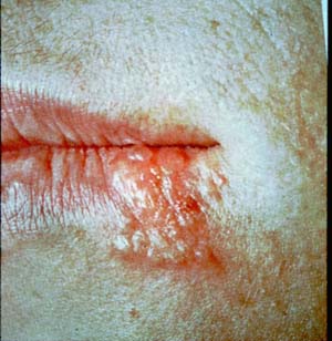
9. Several coalescing vesicles surround by a red halo at
the left commisure the entire lesion is approximately 20x15mm
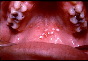
10. Several round, white, firm, raised nodules on the soft
palate, tissue in the center of the lesions appears red and inflamed.
Copyright © 2000
Northern Arizona University
ALL RIGHTS RESERVED