Microscope Notes
The compound microscope is a useful tool for magnifying objects up
to as much as 1000 times their normal size. Using the microscope
takes lots of practice. Follow the procedures below both to get the
best results and to avoid damaging the equipment.
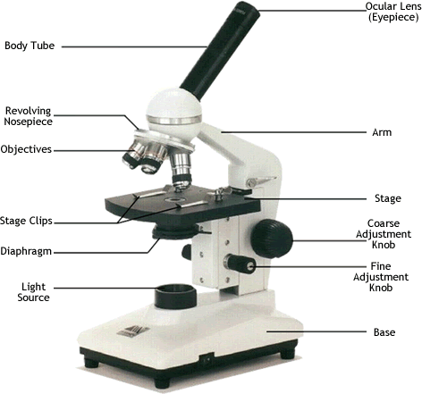
- The eyepiece, also called the ocular lens, is a low power lens.
- The objective lenses of compound microscopes are
parfocal. You do not need to refocus (except for fine adjustment) when switching to a
higher power if the object is in focus on a lower power.
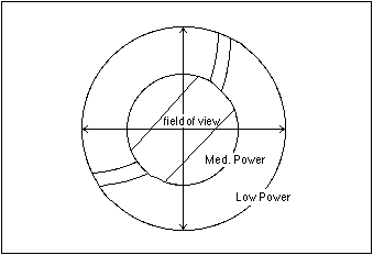
- The field of view is widest on the lowest power objective.
When you switch to a higher power, the field of view is
closes in. You will see more of an object on low power.
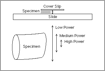
- The depth of focus is greatest on the lowest power
objective. Each time you switch to a higher power, the depth of
focus is reduced. Therefore a smaller part of the specimen is in
focus at higher power.
- The amount of light transmitted to your eye is greatest at the
low power. When you switch to a higher power, light (and therefore
resolving power, or the ability to distinguish two nearby
objects as separate) is reduced. Compensate with the light control
(sometimes called the iris diaphragm).
Field of View
The field of view is largest on the lowest power objective. When
you switch to a higher power, the field of view closes in towards the center. You will
see more of an object on low power. Therefore, it is best to find an
object on low power, center it, and then switch to the next higher power and repeat.
Depth of Focus
The depth of focus is greatest on the lowest power objective. Each
time you switch to a higher power, the depth of focus is reduced.
Therefore a smaller part of the specimen is in focus at higher power.
Again, this makes it easier to find an object on low power, and then
switch to higher power after it is in focus. A common exercise to demonstrate depth of focus involves laying three different colored threads one on top of the other. As the observer focuses down, first the top thread comes into focus, then the middle one, and finally the bottom one. On higer power objectives one may go out of focus as another comes into focus.
Microscope Troubleshooting
Problem #1: The image is upside down and/or backwards.
- Is the slide right-side up?
- Inversion of the image is normal on some microscopes.
- A common demonstration involves looking at the letter "e" on a slide.
- When you move the slide left, does the image move left or right?
Problem #2: Everything is dark.
- Is the microscope plugged in?
- Is the power switch on?
- Is the objective lens snapped into position?
- Is the light control set correctly?
- If you are on the highest power objective, did you forget immersion oil?
Problem #3: I can't find anything on low power!
- Center the coverslip of the slide under the objective lens.
- Focus up and down with the coarse adjustment knob.
Problem #4: When I moved to a higher power, everything
disappeared!
- Return to the previous (lower power) objective.
- Center the object in the field of view.
- Go to the higher power objective and use only the fine focus.
Problem #5: The image is blurry on all powers.
- Clean the microscope's ocular lens. (Only use lens paper!)
- If you rotate the ocular and the specks move, there is dirt on the ocular lens and it should be cleaned.
- Clean the slide. A tissue, paper towel, or cloth can be
used.
Problem #6: The image is blurry only on a particular
power.
- Clean the microscope's objective lens. (Only use lens
paper!)
Microscope Drawings
When drawing what you see under the microscope, follow the format
shown below. It is important to include a figure label and a subject
title above the image. The species name (and common name if there is
one) and the magnification at which you were viewing the object
should be written below the image. All relevant parts of the drawing
should be labelled on the right side of the image using straight
lines. Lines should not cross. Drawings should be done in pencil,
while labels should be in pen or typed. Remember that total
magnification is determined by multiplying the ocular x
objective.
Viewing Prepared Slides
*** Don't hoard slides! You can only view one at a time, so
that's all you should be holding. Return it before getting another,
and if you break it, tell your instructor so that it can be properly
cleaned up and replaced! ***
- Start by rotating the objective lens to lowest power.
- Place a slide on the stage, label side up, with the coverslip
centered.
- On LOW POWER ONLY, use the coarse focus knob to get the
object into focus.
- If you cannot see anything, move the slide slightly while
viewing and focusing.
- If nothing appears, reduce the light and repeat step 4.
- Once in focus on low power, center the object of interest by
moving the slide.
- Rotate the objective to the medium power and adjust the fine
focus only.
- If needed, rotate the objective to the high power and adjust
fine focus only.
Making a Wet Mount (Live
Prep) Slide
- Use a depression slide if possible-it will have a small
indentation that holds fluid.
- Squeeze the air out of the dropper before you put it in
the sample container. (This prevents bubbles from agitating the
contents of the sample bottle.)
- Decide where to put the tip of the dropper-often the best stuff
settles to the bottom!
- While still squeezing the bulb of the dropper, insert the
dropper into the sample container and partially release the pressure
on the bulb. Fluid should rise up slowly. Gently remove the
dropper from the sample container.
- Increase the pressure on the dropper bulb to add a drop (or two
at most) to the depression of the slide. The liquid should not
overflow across the surface.
- If you will be viewing fast moving organisms, you may wish to
add a drop of thickener such as methyl cellulose or "ProtoSlo" to
slow them down by making the fluid more viscous.
- Slowly lay down the cover slip starting at a 45 degree angle
with one edge touching the slide. This helps to prevent air bubbles
from forming under the cover slip.
- Remember that the microscope light is very intense and the
organisms will survive longer on the slide if you turn it off when
not observing.
Further Investigation
Digital microscope for Macintosh or Windows
Investigating pondwater organisms
Powers of 10 (1977 version)
Make your own microscope