SPH405
| Neurological Foundations of Speech, Language and Hearing |
Foundations of Central Auditory Anatomy and Physiology
GOAL: To present the foundations of central auditory anatomy and physiology.
OBJECTIVES: After reading and lecture, the students will:
Describe the extent and general route of the central auditory pathways.
Apply the anatomy and physiology of the central auditory system to the
procedures employed in an audiological evaluation.
Apply the physiology of the central auditory system to the procedures
involved in the speech and language evaluation.
The central auditory system begins at the connection of the
vestibulocochlear nerve (viii) with the brainstem. This connection is at the
cochlear nuclei.
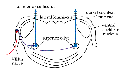
There are two pairs of cochlear nuclei: the ventral cochlear nuclei and the dorsal cochlear nuclei.
Please open the link below and view the position of the ventral cochlear nuclei and dorsal cochlear nuclei in the diagram.
Diagram showing the Cochlear Nuclei
COCHLEAR NUCLEI have Synapses are located roughly where the cerebellum and the pons connect. This area is called, oddly enough, the cerebellopontine junction.
Each fiber of the acoustic branch of the VIIIth nerve divides into
two sections (bifurcates) at this junction.
One branch goes to the dorsal cochlear nucleus,
Another branch goes to the ventral cochlear nucleus.
These branches divide again, sending fibers to anterior
and posterior divisions of the ventral cochlear nuclei
(anteroventral; posteroventral).
Click below for a diagram of the Auditory Pathways.
Diagram of Auditory Pathways
The cochlear nuclei (there are two) are histologically heterogenous. That means their tissues are slightly different.
They contain subdivisions and distinct regions.
These regions represent the TONOTOPIC regions of the organ of corti. Tonotopic regions of theb basilar membrane represent frequencies of sound energy in the audible (for human beings) range from about 20 Hz. to about 20 KHz.
Neuron connections in the cochlear nuclei are arranged to provide a large number of combinations for receiving and contrasting peripheral impulses.
- Each cell of the cochlear nuclei receives terminal endings from about 400 VIIIth verve fibers.
- Each nerve fiber sends terminal endings to about 400 different Cochlear Nucleus cells.
- There are also interneural connections within the Cochlear Nuclei. Interestingly, the retina is arranged the same way.
The SUPERIOR OLIVARY COMPLEX receives fibers from the cochlear nuclei for the transmission of sound sensations.
Please view the diagram in the link below to better understand the role of the superior olivary complex in the auditory pathway.
Full Model of Auditory Pathways
Its location is at the rostral end of a region where many nerve fibers
tracts cross from one side of the body to the other side.
- This is the trapezoid body. The trapezoid body is a large complex of axons and some nuclei. It begins in the lower medulla and reaches rostrally to the caudal pons. Decussation of the pyramidal cells and of the somesthetic posterior column-lemniscal cells occurs here, too.
- About 85% of the nerve fibers cross from one side to the other side, and the rest ascend ipsilaterally. Most, but not all, sound impulses transmitted to the left ear will be sent to the right cerebral cortex.
- Physiologists believe that, for the auditory sense, phase and other temporal (onset) differences that help the localization and/or tracking of acoustic sources are sorted out by decussation at the trapezoid.
- The Superior Olive receives fibers from the anterior part of the ventral cochlear nucleus, but not from the other parts. The rest of the fibers, as well as the ones that synapsed at the superior olive travel rostrally along the brainstem from the pons to the midbrain.
Nerve impulses also originate in the brainstem and propagate to the
cochlea.

These efferent potentials are fired through fibers that originate at the superior olive. The purpose of these impulses is not clear.
The tract is called the olivocochlear bundle fiber tract. Audiologists now can measure otoacoustic emmissions in subjects who can not or will not allow a reliable threshold determination.
The inferior colliculus of the midbrain is the next site for a synapse of central auditory fibers. (Do you recall the name of the midbrain synapse site for visual fibers?)
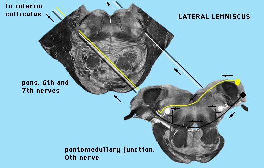
Not all of the fibers synapse here, but they all continue on to the thalamus along the LATERAL LEMNISCUS.

Again, some fibers appear to synapse along this tract at the NUCLEUS of the LATERAL LEMNISCUS.
While others push on rostrally.
At the Inferior Colliculus, there remains a great deal of confusion about the exact anatomical arrangement of nerve fibers. In fact, rostral to the Superior Olivary Complex, fibers are packed to tight and are so interconnected that it is very difficult to sort out and trace impulses.
There is speculation that connections of the ears to the mm. that control the eyes exist here. This makes some sense for those of us who recall that the fibers from the optic tracts synapse here on their way to the pretectal nucleus of the superior colliculus to control the light reflex.
Such a connection might serve to reflexively turn an animal's (your) gaze toward the source of a sound.
We might also include the muscles that turn the head or blink the eyes in this reflex arrangement.
The Thalamus is the next big structure of the brain through which the sound generated impulses travel.

The thalamus is the colored purple in the image above.
The thalamus receives all sensory input bound for the cerebral cortex.
The medial geniculate body is the place for hearing in the thalamus.
Each sense has a special place in the thalamus.
At this time there is little convincing evidence of a tonotopic arrangement in the Medial Geniculate Body of the Thalamus.
The "Auditory Cortex" is the last stop for sound generated nerve impulses.
In human beings and other primates, the auditory cortex is
located on the superior temporal gyrus.
Within the Fissure of Sylvius in each hemisphere of the
brain, is an area called Heschl's Gyrus.
The surrounding cortex is auditory association cortex.
The auditory cortex of each hemisphere receives input from each ear.
There is some evidence to suggest that there is, in most subjects, a right ear preference for certain auditory events and a left ear preference for others.
These findings correspond with studies that show a left hemisphere superiority for consonants and other segmental aspects of speech while...
There appears to be a right hemisphere preference for vowels, suprasegmental features and music.
The temporal lobes of the cerebral cortex are the locations for the processing of speech.
In general, we speak of differences in the functions of the anterior versus the posterior portions of the temporal lobes.
Studies of living subjects reported activity of the superior gyri of the temporal lobes upon presentation of sound stimuli. (Penfield, W., and Roberts, L. (1959). Speech and Brain Mechanisms. Princeton: Princeton UP.; Ojemann, G. (1991). Cortical organization of language. Journal of Neuroscience, 11, 2281-2287).
The areas around the primary auditory cortex deal with the
interpretation of sound impulses. These areas are called the
association areas.
Further to the posterior, and spanning into the parietal lobe of
the dominant hemisphere, the cortex performs the complex task
of linguistic symbolic decoding and encoding.
The auditory evoked response is now a standard means of examining central auditory system function.
Surface electrodes are placed on the scalp, usually at the vertex of the skull.
A stimulus, usually a "click" or some other transient aperiodic sound, is presented through headphones to an ear or to both ears.
The stimulus is presented many times, and the results are summed. The sums are automatically recorded on a strip of paper. This recording paper is calibrated to display the measurement of time elapsed between stimulus presentation and the electrical activity associated with synaptic junction excitation. Examiners infer recorded "peaks " of electrical activity are created when the action potential crosses synaptic clefts at the sites along the auditory pathways: cochlear Nuclei; Superior Olivary Complex; Lateral Lemniscal Nucleus; Inferior Colliculus, Medial Geniculate Body.
It can take up to 300 msec. for a stimulus-generated action potential, called the Auditory Evoked Response or AER, to reach the brain. Naturally, it takes less time for the potentials to reach more caudal structures. AER's recorded at the lower levels of the central system are called BSER's (Brainstem Evoked Responses).
Once you have finished you should:
Go on to Online Lesson 2
or
Go back to Auditory System
E-mail Bill Culbertson
at bill.culbertson@nau.edu
Call Bill Culbertson
at (520) 523-7440
Copyright © 1999
Northern Arizona University
ALL RIGHTS RESERVED