SPH405
| Neurological Foundations of Speech, Language and Hearing |
Form and Function of the Peripheral Visual Mechanism
GOAL: To relate the form and function of the peripheral visual mechanism to normal human communication.

OBJECTIVES: After reading, lecture and study, the students will:
Apply the A/P of the peripheral visual mechanism to evaluation of the human communication system.
Name the extra ocular m.m. and their actions
Describe three effects of age and disease on the peripheral vis. mechanism.
Differentiate the accommodation and light reflexes. Explain what these reflexes are and how to observe them.
The visual system is one of the most highly developed systems in man.
In man, vision is a primary modality of communication. for communication, we use vision in gesturing, facial expression and reading/writing.
Vision is highly developed compared to many other animals. Dogs, for instance, have very poor vision. Deer won't win any awards for their visual acuity, either.
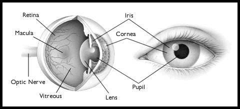
The peripheral visual mechanism consists of the EYEBALL, the OPTIC TRACTS the EXTRA OCULAR EYE MUSCLES, the EYELIDS and the LACRIMAL SYSTEM.
The eyeball is the organ of sight.

It converts diffuse photic energy into neural impulses (action potentials).
It consists of e FOCUSING MECHANISM, and a system of PHOTORECEPTORS.
The FOCUSING MECHANISM is the IRIS, CORNEA and LENS. The iris helps modify the amount of light that reaches the retina. It opens to allow more light to reach the PHOTORECEPTORS of the retina.
Open=dilate; close=contraction (myosis)
The pupil closes for two reasons. It helps focus distant objects on the retina. It shades the retina from excessive light energy.
Several intrinsic eye muscles adjust the iris. the receive their peripheral innervation from the oculomotor nerve (III):

The CIRCULAR MUSCLE contracts and causes the sphincter pupillae to constrict.
The RADIAL MUSCLE causes dilation of the pupillae.
The cornea REFRACTS the light to focus it on the retina. Naturally, the better focusing we have, the clearer is our picture.
The Cornea is the most anterior part of the eyeball. It is made of ectoderm.
Embryonically, it arises from the dura.
The Cornea provides for most of the light focusing.
In man, it focuses twice that of the lens. If is anterior surface becomes irregular, astigmatism is the result. The lens fine tunes the focus of light to the retina.
This fine tuning is called ACCOMMODATION.
It is controlled by the oculomotor nerve (III).
The ciliary muscle flattens the lens for accommodation.
It relaxes tension on the lens by tightening the anterior section of the eyeball
The lens becomes more concave or round when the ciliary muscle relaxes.
With age, the tissue of the lense gets less flexible, and the lens fails to return to its round state.
This causes difficulty seeing near objects or HYPERMETROPIA.
If the individual has difficulty seeing distant objects, the condition is called MYOPIA
With age, the lens also becomes more dense and opaque. This condition is called
a "CATARACT". It can be corrected surgically
The PHOTORECEPTOR SYSTEM is contained in the RETINA.
Photosensitive receptors on the surface of the retina convert light to neural impulses.
Light is projected as an inverted, backwards pattern on the surface of the retina.
The receptors are RODS and CONES.
Rods (around 120 million of them) are found in the peripheral part of the retina. They are more light sensitive...but they are less color sensitive.
Cones are color sensitive. There are fewer of them (7 million. They are concentrated in the center of the FOVEA CENTRALIS. The fovea centralis is the functional center of the retina and area for greatest visual acuity.
The retina occupies the FUNDUS of the eye. Funduscopic examination is performed by the MD. It reveals pathological conditions
There are (normally) two eyeballs:
OD (oculus dexter ) means RIGHT EYE.
OS (oculus sinister ) means LEFT EYE.
Two eyes allows for BINOCULAR VISION. This helps us have perspective and perception of depth. It also allows redundancy of the system
The eyeballs are contained in the orbits.
The orbits are located below the frontal lobes, lateral to the nose.
The orbits contain other structures associated with vision. Lacrimal glands secrete tears. Extra ocular muscles orient the eyes. Conjunctiva provides a tissue matrix for the eyeballs. The optic nerves convey light generated action potentials to the central nervous system.
The OPTIC TRACTS convey neural impulses to be interpreted as sight from the photoreceptor system to the cerebral cortex and to other neural structures.
These tracts begin at the retina, collect in the twin OPTIC N.N. (II: actually a tract rather than a nerve.
They optic tracts converge in the OPTIC CHIASMA
There is a mixed decussation here. Nasal fibers cross. Inferior fibers loop.
These fibers follow divergent courses and terminate in various centers of the diencephalon and mesencephalon.
Fibers bound for the visual cortex terminate in the LATERAL GENICULATE NUCLEUS of the thalamus.
Some terminate in the mesencephalon via the PRETECTAL NUCLEUS and the SUPERIOR COLLICULUS.
These fibers transmit impulses concerned with the light reflex and the accommodation reflex.
The EXTRA OCULAR EYE MUSCLES control the direction of ocular gaze.
When we talk about eye movement or parts of the eyeball, anatomy we refer to locations based on the face.
Nasal is toward the nose.
Temporal gaze is lateral.
When an eyeball is fixed toward the nasal area, the condition is said to be esophoria.
The opposite condition is exophoria.
There are 6 extra ocular eye muscles
1. Medial Rectus: Moves eye temporal medial
2. Superior Rectus: up/nasal
3. Lateral Rectus: to temporal lateral
4. Inferior Rectus: nasal/ down
5. Superior Oblique: temporal /down
6. Inferior Oblique: temporal/ up
These muscles are innervated by III, IV and VI
All send afferent fibers to midbrain to coordinate contractions.
The eyelids CLOSE OFF LIGHT TO THE RETINAE.
The lids are CLOSED by orbicularis oculi
These muscles are part of the muscles of facial expression They receive their motor innervation from the facial nerve (VII).
This closing allows for more restful sleep
It also allows for lubrication of the eyeball.
The lids are Opened by LEVATOR PALPEBRAE SUPERIORIS (III)
Some individuals have difficulty opening the eyelids. This condition is called Ptosis.
Difficulty closing the eyelids can cause extreme discomfort.
The LACRIMAL SYSTEM consists of the lacrimal glands and the tear ducts
Lacrimal glands secrete tears. Tears have several functions. They lubricate the eyes. The display emotion: laughing and crying.
Tear ducts convey tears to eye from the lacrimal glands.
Once you have finished you should:
Go on to Online Lesson 2
or
Go back to Visual System
E-mail Bill Culbertson
at bill.culbertson@nau.edu
Call Bill Culbertson
at (520) 523-7440
Copyright © 1999
Northern Arizona University
ALL RIGHTS RESERVED