SPH405
| Neurological Foundations of Speech, Language and Hearing |
![]()
![]()
![]()
![]()
![]()
![]()
The Corticobulbar Tracts of the Pyramidal System
GOAL: TO PRESENT THE CORTICOBULBAR TRACTS OF PYRAMIDAL SYSTEM
OBJECTIVES:
Differentiate the structures and functions of the divisions of the pyramidal tracts.
Associate the functions of the corticobulbar tracts with communication and swallowing.
Trace the corticobulbar tracts from the frontal lobe to the bulbar musculature.
The PYRAMIDAL SYSTEM Is the neurological structure by which voluntary, conscious, motor impulses travel from the frontal lobe to the muscle the individual intents to contract.
Corticobulbar tract #2 feeds into the motor nuclei of cranial nerves V; VII; X-XI; XII.
Corticospinal tracts are for spinal motor nerves. spinal motor nerves emerge from the ventral aspect of the cord. with final common synapses in the anterior gray horns.
Corticobulbar #1 pathway begins in the cerebral cortex.
Upper Motor Neuron cell bodies lie here.
Axons go to INTERNAL CAPSULE.
They form a compact bundle and enter at the genu, which looks like the knee of a little leg.
They are bound for the motor nuclei of to III IV & VI.
This gives bilateral innervation for the muscles served by III, including the superior rectus; medial rectus; inferior rectus and inferior oblique; and the levator palpebrae superioris.
Cranial nerve III leaves the brainstem at the upper midbrain level.
IV fibers aren't crossed, and descend ipsilaterally to the nucleus of the trochlear nerves.
The anatomical arrangement of IV is distinctive: it is the only Cranial nerve to leave the brainstem from the dorsal aspect.
IV supplies motor impulses to the superior oblique muscles to allow eye gaze temporally and interiorly.
There are ABERRANT fibers of IV that join up with VI for coordination of eye muscles.
Fibers of VI are all crossed before they reach the nucleus of VI
To reach the lateral rectus muscles which rotates the eye to the temporal aspect.
Cortocobulbar #2 fibers provide motor innervation to the motor nuclei of cranial nerves V, VII, IX, X-XI and XII.
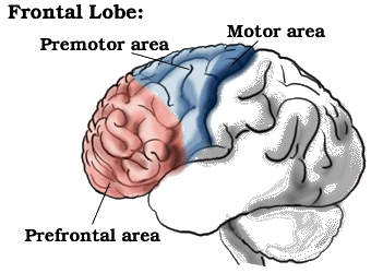
They originate in the lateral 1/3 of the precentral gyrus of the frontal lobe: area #6.
Axons course from here to the genu of the internal capsule.
- Tracts that innervate motor nucleus of VII for the lower muscles facial expression.
- Those that go to the portion of X-XI for innervation of the levator Veli Palatini muscle
- Those that supply the XII for muscles tongue.
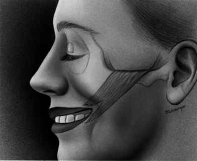
This means that lesions of the right Upper Motor Neurons will result in
- Paralysis of the left lower muscles facial expression. The patient will smile emotionally, but not voluntarily.
- Paralysis of the left levator Veli Palatini (velum will move up and right).
- Paralysis of the left tongue muscles (the tongue will protrude to right).
Once you have finished you should:
Go on to Online lesson 2
or
Go back to Major Descending Tracts
E-mail Bill Culbertson
at bill.culbertson@nau.edu
Call Bill Culbertson
at (520) 523-7440
Copyright © 1999
Northern Arizona University
ALL RIGHTS RESERVED