SPH405
| Neurological Foundations of Speech, Language and Hearing |
![]()
![]()
![]()
![]()
![]()
![]()
Development of the Central Nervous System
GOAL: To establish an understanding of development of the human nervous system upon
which to base management of communicative disorders.
OBJECTIVES: "After reading and lecture, the students will..."
Describe the development of the human nervous system, including the
following high points:
- Formation of the neural tube
- Differentiation of the neural tube, including the primary brain
vesicles and the spinal cord.
- Derivative structures and cavities of the prosencephalon,
telencephalon, diencephalon, rhombencephalon and medulla
spinalis.
Describe the development of the cerebral hemispheres.
... on the section examination with 90% accuracy.
(FOR SUPPORTING TEXT, see: Zemlin, W. (1988) Speech and Hearing Science: Anatomy
and Physiology (3rd. Ed.). Englewood Cliffs, NJ: Prentice-Hall Ch, 7 and Clarke, W. Central
Nervous System in Hamilton, W. (1976). Textbook of Human Anatomy. St. Louis, MO: C.V.
Mosby).
Embryological Development of the Human Brain
General Overview
All the events that occur during nervous system development are fascinating, but are
sometimes difficult for the beginning student to grasp. We will focus on three major
events that occur in the development of the human nervous system: development of the
neural tube; differentiation of the neural tube into the primary brain vesicles; and the
development of the cerebral hemispheres.
Development of the Central Nervous System (Overview with diagrams)
You should remember, then, that this lecture is a great simplification of the many
events that actually occur. Many more events occur, and vary, depending upon the
genetic foundation of each individual. A few of the many significant mileposts that
we are just "blowing past" include:
The appearance of the Neural Folds on either side of the Primitive Streak.
The Streak is the first sign that ectodermal cells are differentiating into the
complex structure that will eventually become the human nervous system.
- The development of differentiated cells that will form the functional units of the nervous system, which we shall come to know as Neurons: The Neuroblasts.
- The migration of the Peripheral Nerves from the Neuromeres of the primitive spinal cord to establish Dermatomes and Myotomes. Dermatomes are regions of sensation on the body. Myotomes are regions of muscle function on the body. These regions coincide, and correspond to segments of the spinal cord. Dermatomes evolve as the embryo develops along lines established by developing blood vessels, connective tissue and muscle planes.
But our current goal is to examine neurological embryology ("Neuroembryology")
for the purposes of case management in speech-language pathology. We'll just
get overwhelmed if we don't focus. While it is difficult to hold that any embryonic
event is insignificant with regards to the overall functioning of the individuals we
will treat, we will keep things focused on a few critical events.
- These events bear direct import to the success of communicative development or to understanding the anatomy and physiology of the Nervous System.
- There is an interplay of endogenous and exogenous factors which will modify the outcome of the embryonic development of the Nervous System.
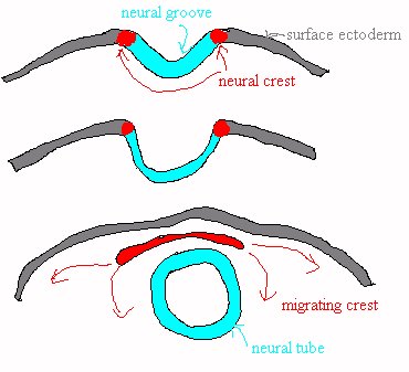 The NEURAL TUBE is created when the developing embryo, itself forming in the shape
of a tube, forms a groove along the axis of its dorsal surface. The embryo has already
begun to form three different types of cells as it enlarges. The outer cells are ectodermal
cells. Since the neural groove starts in the surface of the embryo, its cells will be formed
of ectoderm, folded into the interior of the embryo.
The NEURAL TUBE is created when the developing embryo, itself forming in the shape
of a tube, forms a groove along the axis of its dorsal surface. The embryo has already
begun to form three different types of cells as it enlarges. The outer cells are ectodermal
cells. Since the neural groove starts in the surface of the embryo, its cells will be formed
of ectoderm, folded into the interior of the embryo.
This folding occurs between the 16th and the 28th day after fertilization. At about the time there are 19 somites. Somites are blocks of tissue-forming segments along the length of the embryo. Embryologists like to refer to embryonic developmental stages in terms of how many somites there are, since somite appearances are a more reliable mileposts in the chronology of embryonic development than are calendar days. Embryos don't look at the calendar, you see, but most of them seem to form somites in a regular pattern. There will be a total of about 30 somites by the end of the fourth week, depending upon the individual.
The primitive tube is open at each end. These openings will eventually close. The rostral opening is the Anterior Neuropore. The caudal opening is the Posterior Neuropore. After the tube fuses, closing the ends, mesoderm covers its outside. Then, more ectoderm covers that mesoderm. Thus, the ectoderm that forms the nervous system has become enveloped into the interior of the embryo. Ectodermal disorders may be noticed on the skin as well as the nervous system.
The neural tube will eventually become the entire nervous system, including both central and peripheral divisions. As growth proceeds, Neural Tube cells differentiate and give rise to the all cells of the nervous system. At first, all the neural tube cells are Medullary Epithelial cells, formed from the ectodermal germ layer. The different cells of the nervous system result from migrations of the nuclei and cytoplasm of these cells.
The neural tube is thicker on the sides that on the ventral and dorsal surfaces. The cells of the lateral walls of the tube evolve into three layers: the Ependymal zone, the mantle zone and the marginal zone.
Ependymal Cells form the lining of the Nervous System. This, in the adult, will be the interior of the Cerebral Ventricles and the central canal of the Spinal Cord.
The Mantle Zone is the bulk of the nervous system. It is created
by the differentiation of Germinal Cells. Its cells evolve into
Neurons and Glial (support) Cells. The Mantle Zone will become
the gray matter of the nervous system. Gray Matter develops from
several types of Germinal Cells, including:
- Spongioblasts give rise to Astrocytes.
- Medulloblasts form Astrocytes or Oligodendrocytes.
- Astrocytes are support cells. They form a lattice to hold the neurons in a functional structure.
- Oligodendrocytes form myelin, a sheath that insulates the axons and enhances the propagation of action potentials.
- Glioblasts develop into Glial Cells or Neuroblasts.
- Glial cells provide further structure for the nervous system.
- Neuroblasts develop into neurons.
Marginal Zone cells cover the outer surface of the tube. Early in development, Marginal Zone cells are Spongioblasts, providing support for the neural pathways of the maturing Mantle Zone with their multiple protoplasmic projections. Later, neurons will grow up and project long whitish (myelinated) axons into the Marginal Zone. The Marginal Zone becomes the White Matter of the nervous system.
As the tube develops more, ventral (basal) and dorsal (alar) regions differentiate. These regions along the axis of the tube are thinner and flatter than the lateral aspects. Different patterns in their ontogenies (development) are significant in the future function of the nervous system.
-
The basal lamina cells become motor or efferent in function and the
alar laminar cells evolve into sensory or afferent cells. This is a
general trend, most noticeable in the spinal cord, but generally lost
in the brain.
Afferent impulses convey sensation form the body wall and muscles (somatic) and from the internal organs (visceral).
Efferent impulses may be to the internal organs (visceral) or to the body wall and voluntary muscles (somatic).
This is an early phenomenon, occurring before the pores close.
The Anterior Neuropore closes at the 20 somite stage.
The Posterior Neuropore seals at the 25 somite stage.
- The PROSENCEPHALON ("forward brain") will eventually give rise to the cerebral hemispheres and the thalamus.
- Projections of neural tube tissue (diverticula) fold inward into the prosencephalon.
- They divide the prosencephalon into rostral and caudal sections.
- The rostral section is the Telencephalon ("End Brain").
- The Telencephalon will develop into the structures that comprise the Cerebral Hemispheres, including...
- The Cerebral Cortex
- The Basal Ganglia
- It still has a hollow center, even after all this folding, and as it folds into two sections, the hollow parts become the Lateral Ventricles (and part of the Third Ventricle).
- The caudal portion is the Diencephalon ("Dual Brain").
- The diencephalon will develop into the Thalamus and Hypothalamus.
- It is hollow, too. Its cavity is the third ventricle. The Third Ventricle is squashed flat by the developing cerebral hemispheres.
- The Mesencephalon ("Midbrain") does not differentiate much from the pattern of the spinal cord from which it rises.
- It will become part of the Cerebellum.
- And will give rise to some structures of the rostral part of the brainstem, the midbrain. including:
- The Inferior Colliculus
- Red and Black Nuclei
- Neural tracts develop that connect this part of the brain with the other parts of the brain.
- The Rhombencephalon has two subdivisions: the Myelencephalon and the Metencephalon.
- The Myelencephalon will develop into the medulla oblongata.
- The Medulla Oblongata forms the caudal part of the brain, connecting directly to the spinal cord (Medulla Spinalis).
- From this structure the nuclei of the cranial nerves devoted to mastication, facial expression and phonation develop (CN's V-XII).
- The Metencephalon will develop into the Pons and the Cerebellum.
- The Pons houses the major tracts that pass to and from the Cerebellum.
- The Cerebellum coordinates body movement.
 (maf copyright)
Development of the Brain Vesicles is essentially a widening of the tube into three primary regions. Each wide region is a vesicle. Later, the first and the last Vesicles subdivide, resulting in a total of five vesicles.
The first three vesicles to develop are the Prosencephalon, the Mesencephalon
and the Rhombencephalon.
- These cells accumulate and will become the gray matter of the hemispheres: the cerebral cortex. The cerebral cortex is the thin layer of neurons (and glial cells) that covers the surface of the cerebral hemispheres.
- The axons of neurons derived from mantle cells become myelinated and pass into the substance of the brain. These axons are to become the white matter of the cerebral hemispheres.
- As the fetal Telencephalon develops, a deep separation appears along the longitudinal line of the rostral tube.
- This LONGITUDINAL FISSURE will become the fissure that separates the right and left cerebral hemispheres. it is also known as the Interhemispheric Fissure.
- A LATERAL FISSURE forms as the growth of the outer cortex of the hemisphere overtakes the slower growth of the underlying tissue (corpus striatum). These outer cortical tissues fold over, and cover part of the cortex. this process is called "Operculation." Some associate the degree of operculation with intelligence.
- The part of the cortex that surrounds the Lateral Fissure is called the Opercula.
- The part of the cerebral cortex that is covered by the opercula is called the Insula.
- There are FOUR PARTS of a cerebral hemisphere
- The Cerebral Cortex is the thin outer coating of each hemisphere. Here is the seat of the highest level of mental processing.
- It is composed of relatively slow firing neurons, without myelination. This is part of the so-called "Gray Matter" of the brain.
- It grows very rapidly, out-pacing the growth of the skull in which it is housed. Rapid growth causes wrinkled appearance (CONVOLUTIONS) at about 4th month in utero.
- The cerebral hemispheres completely cover the diencephalon and mesencephalon by the 5th month
- The Rhinencephalon ("nose brain") is the rostral part of the brain underlying the cerebral cortex. It is also called Archipallium.
- Develops into the primitive brain structures associated with instinctive behavior.
- These include the Olfactory lobes and the Hippocampus
- The Corpus Striatum serves as a relay system for major ascending and descending neural pathways. There are several components of the corpus striatum, and different anatomy texts will group these components differently. They include:
- The Internal Capsule
- Caudate Body (curved around the lateral ventricle)
- Lentiform Nuclei (located lateral to the thalamus)
- The White Matter Formation of the cerebral hemispheres is composed of the axons of the gray matter neurons at the surface. These axons lie between the cortex and the corpus striatum.
- They enable the brain to send and receive impulses to and from more caudal parts of the central nervous system and across adjacent parts of the brain.
- Since they derive from the development of the cortex, their growth occurs at a rapid rate.
Once you have finished you should:
Go on to Online
Lesson 2
or
Go back to Central Nervous System I
E-mail Bill Culbertson
at bill.culbertson@nau.edu
Call Bill Culbertson
at (520) 523-7440
Copyright © 1999
Northern Arizona University
ALL RIGHTS RESERVED