SPH405
| Neurological Foundations of Speech, Language and Hearing |
![]()
![]()
![]()
![]()
![]()
![]()
Gross Anatomy of the Brain
GOAL: To introduce the gross anatomy of the brain.
OBJECTIVES: After Lecture and Reading, the students will be able to:
Describe the gross anatomy of the brain.
Locate and state the general functions of :
- The Telencephalon
- The Diencephalon (Thalamus and Hypothalamus)
- The Brainstem
- The Cerebellum
Relate the functions of the cerebral hemispheres, thalamus, brainstem and
cerebellum.
... on the examination @ 90%.
At this point, we have discussed the rationale for studying Neuroscience as it applies to human
communication, and we examined the general organization of the human Nervous System. Our
first attempt at organizing the human Nervous System was to separate it into two main divisions:
the Central Nervous System and the Peripheral Nervous System. Now we are going to examine
the Central Division in closer detail, beginning with the Brain. But first, let's review:
REVIEW OF THE CENTRAL NERVOUS SYSTEM
- As you recall from the "General Organization Lecture" the CENTRAL NERVOUS SYSTEM is composed of the BRAIN and the SPINAL CORD.
- It is made up of a mass of nerve fibers (neurons) embedded in a matrix of connective tissue (neuroglia) and coated in three layers of nutritive and protective tissue (meninges).
- The CNS is contained in the bones of the Cranium and Spinal Column.
- We are going to focus on the Brain, which, as you might guess, is housed in the Cranium.
- The Central Nervous System is functionally a mediator between the receptors and effectors of the PNS.
- It controls, regulates and initiates all cognitive and sensorimotor functions. (Question: what behavior does the CNS initiate ?)
- The highest and most intricate mediator for the PNS is the Brain. We've spent a of time evolving and developing this structure. We probably have a long way to go...
- The BRAIN is composed similar halves called The Cerebral Hemispheres, interconnecting fibers, a central structure, composed of the Thalamus and Hypothalamus, a Brainstem, and two hemispheres of the Cerebellum.
- THREE PARTS of the brain form during embryonic development. These parts will develop into the structures we see in the adult brain.
- The three embryonic parts are the Prosencephalon ("Forward Brain"), Mesencephalon ("Middle Brain"), and Rhombencephalon ("Part of the Brain That Looks like an Oblique Parallelogram")..
- The three parts, called Primary Brain Vesicles, form at the rostral end of the Neural Tube as it enlarges and tissues begin to diversify.
- This is the first main division of the Nervous System.
- The Prosencephalon divides again in later development.
- It forms into the Telencephalon ("End Brain") and the Diencephalon ("Twice Brain").
- The Telencephalon will eventually form the Cerebral Hemispheres, including:
- Cerebral Cortex.
- Basal Nuclei (Ganglia)
- It will retain its hollow center, which will develop into two halves called Lateral Ventricles.
- The Diencephalon will form the Thalamus, and the Hypothalamus.
- It will retain a hollow center, too.
- The center will become known as the Third Ventricle.
- The two CEREBRAL HEMISPHERES are structurally and functionally similar.
- Each is composed of several layers.
- An thin outer layer of Cerebral Cortex.
- Deep, inner layers called SUBCORTICAL structures, and ...
- An intermediate mass of "white matter" fibers,
- The Cerebral Cortex communicates with the subcortical structures by means of the intermediate white (myelinated) fibers.
- It also communicates with other areas of the cortex via these fibers.
- The Cerebral Hemispheres serve a wide range of functions.
- They receive, bring to consciousness, coordinate, integrate, interpret and store and retrieve input from the environment.
- They are the initiators of voluntary movement.
- The Cerebral Hemispheres form the most visible part of the human brain.
- They are located on either side of the Interhemispheric (Longitudinal) Fissure.
- They surround the Lateral Ventricles.
- The CEREBRAL CORTEX is the outer layer of the Cerebral Hemispheres.
- Anatomists identify two (or maybe three) parts of the Cerebral Cortex:
- The NEO (new) CORTEX, which is the seat of the most phylogenetically advanced (evolved) functions of the brain.
- Its functions are RECEPTIVE; ASSOCIATIVE; INTEGRATIVE; AND PROJECTIVE.
- The Neocortex is the part of the brain most readily identified with conscious communicative intentions.
- In addition to the neocortex, there are the ARCHI (old) CORTEX and the PALEO (ancient) CORTEX.
- These special types of cortex are found at the medial areas of the temporal lobes.
- Paleocortex is located at the medial aspect of the temporal lobe, roughly in the center (area 28, the Entorhinal area. Remember the "Rhinencephalon" or "Nose Brain?").
- Archicortex is located at the area called the HIPPOCAMPUS.
- Why these areas have their separate semantically identifying names is unclear, because they appear as cortexes at about the same level of (phylogenetic) evolution: reptiles.
- The thickness of the Cerebral Cortex ranges from 1.5 to 4.5 mm. The tissue of the cortex is wrinkled, allowing it to crowd more thinking cells (neurons) into the skull. The wrinkles are called CONVOLUTIONS. Elevated parts of convolutions are GYRI (singular: gyrus), and depressed parts are SULCI (sing.: sulcus. A particularly important or large sulcus is called a FISSURE.
- Another characteristic of the cortex is its STRATIFICATION. It is arranged in several layers, like a cortical sandwich.
- Some anatomists identify 6 layers of tissue with different names according to different anatomists.
- Molecular level
- External granular layer
- Pyramidal layer
- Internal granular layer
- Ganglion cell layer
- Multiform layer
- These six layers can be simplified (LeGros Clark in Hamilton, 1976) into four strata.
- Zonal Layer.
- At the surface
- Horizontal fibers
- Few and scattered cells
- Supragranular Layer (Pyramidal Layer)
- This is the thickest layer.
- It has closely packed, pyramid-shaped cells
- Granular Layer: comprised of small round cells with short axons and dendrites
- Infragranular Layer: comprised of cells of several types
- CONNECTIONS of the CEREBRAL CORTEX: The Neurons of the Cerebral Cortex are connected in different ways, depending upon their functions.
- Some have long axons traversing the white matter and beyond.
- An example of these are the Pyramidal Cells of the voluntary motor tracts.
- These send axons into the Brainstem and on down into the Spinal Cord.
- Others project long axons horizontally to other neurons at distant cortical locations.
- These are the Association Fibers, which relay information to and/or from various neural centers.
- One long fiber bundle, the Arcuate Fasciculus, extends from the frontal pole to the parietal area.
- Another, called the Uncinate Fasciculus, extends from the posterior parietal area to the Temporal Lobe area.
- This suggests the Temporal Lobe receives input and sends output from and to other parts of the brain.
- This fact has important function for integration of the cortex as a whole.
- Some cortical neurons have short axons to communicate with other cells in the same region at near locations (from gyrus to gyrus).
- Some deep neurons send axons to neurons in more superficial layers.
- INPUT to the CEREBRAL CORTEX: Neurons of the Cerebral Cortex receive input from various sources.
- Thalamus.
- Contralateral hemisphere.
- Ipsilateral hemisphere.
- Neighboring neurons.
- FUNCTIONAL AREAS of the CEREBRAL CORTEX: Specific areas of the cortex have been identified with specific functions.
- These are meeting points for certain kinds of input from special centers of the CNS.
- Damage to these areas (through neoplasm, vascular damage, trauma, etc.) produces certain signs/symptoms.
- In 1955, Brodmann experimented with the brain, and identified important areas associated with certain functions. He numbered them, as you see below. There are many more numbered areas not included in this lecture, We will examine the areas of greatest significance to communication science, according to the "Lobe" in which they are located.
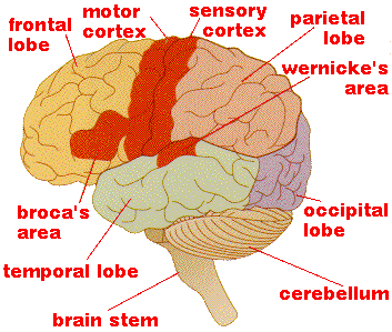
LOBES of the CEREBRAL CORTEX: The Cerebral cortex is divided into Frontal, Parietal, Temporal and Occipital Lobes. These lobes are named after the bones of the Calvaria under which they lie: FRONTAL; PARIETAL; TEMPORAL and OCCIPITAL. Bearing in mind that the Brain FUNCTIONS BEST AS A WHOLE (in concert), physiologists have attributed functional roles to the lobes. These functions may be described as "primary" either or "association."
FRONTAL LOBE: The primary functions of the frontal lobe appear to be motor and intellectual. The Frontal Lobe Lies beneath the frontal b. There are several functions associated with the Frontal Lobe, including...
Motor Cortex (Precentral Gyrus): Area #4 and Area #6: Precentral and
Paracentral Gyri. Here are located the cell bodies of the pyramidal tracts:
Corticospinal and Corticobulbar. As their names imply, the Pyramidal
Tracts extend from the Cerebral Cortex to the Spinal Cord or to the Bulb
(Brainstem). A lesion of the cortical motor strip results in paralysis or
paresis. The cortex has a function of inhibition as well as initiation of
motor impulses.
- The Precentral Gyrus is called the PRIMARY MOTOR STRIP. It is the location of the cell bodies of the Corticospinal Tracts, which provide innervation to the axial and appendicular body below the neck, and the Corticobulbar #2 Tracts, which innervate all head and neck musculature except the muscles of the eyes. Thus, when an individual is affected by damage to the motor cortex, he will experience degrees of inability to initiate movement, and the stretch reflex will not be inhibited.
- Homunculus (Projections): The motor cortex is arranged in a SOMATOTOPIC pattern. Traditionally, anatomists like to draw a little man stretched out along the motor strip. This little guy is called the "Homunculus."
- We can stimulate areas associated with this little man and the same areas on the body will respond. For example, if we stimulate the imaginary leg of the Homunculus, the contralateral leg on the patent's body will respond.
- Area #6 is the "Paracentral"Gyrus, "Voluntary Eye Field": the posterior aspect of the middle frontal gyrus. It is the site of the cell bodies of the Corticobulbar #1 tracts, innervating the eye muscles.
Areas #9. #10, #11, #12: is called the Prefrontal Cortex. It is associated with integration of many of the fibers in the long association bundles. (See Ojemann and Mateer, 1979). Damage to these areas affects affective behavior, including memory and emotion.
PARIETAL LOBE: The Parietal Lobe is generally associated with tactile
reception and interpretation. The tactile sense includes the awareness of physical
contact as well as the sensation of position and movement. The Parietal Lobe lies
beneath the parietal bone, Posterior to the Frontal Lobe; superior to the Temporal
Lobe; anterior to the Occipital Lobe.
- SOMESTHETIC CORTEX Areas # 3, 1 and 2: Posterior to the Central Fissure is the Somesthetic Cortex, associated with tactile reception to consciousness. There is a difference between conscious and unconscious sensory input and the Postcentral strip receives conscious tactile sensations. Neurons of the Somesthetic Cortex are arranged somatotopically, and correspond to the motor strip in somatotopic representation. They serve, generally, the function of bringing sensations of pain, temperature, gross touch, vibratory sense, proprioception to consciousness.
- SOMESTHETIC ASSOCIATION Areas #5 &7 are associated with Stereognosis: the ability to recognize objects by feeling them. They may be considered "Association Areas" of the Somesthetic Cortex.
- SYMBOLIC FUNCTIONS: #39 and #40 are the Angular Gyrus and Supramarginal Gyrus, respectively. These areas are heavily connected to Broca's Area in the Frontal Lobe and are concerned with articulate speech. Association of auditory (Supramarginal) and visual (Angular) symbols with their referents appears to be seated here. Note that areas #39 and #40 correspond to what we call "Wernicke's Area," and are located at the junction of the Parietal, Occipital and Temporal Lobes.
OCCIPITAL LOBE: The Occipital Lobe is primarily associated with reception
and association of visual impulses. It is located in the posterior portion of the
Brain, deep to the Occipital Bone.
- The PARIETO-OCCIPITAL FISSURE separates the Occipital Lobe from the Parietal Lobe. It is difficult to distinguish the borders of the Occipital Lobe by examining the Brain's lateral surface. However, if you look at the medial surface of the hemisphere, the Parieto-Occipital sulcus is clearly visible. This Sulcus separates the parietal and occipital lobes.
- Visual Input Functions of the brain are processed at the same levels as are Auditory functions or any other special sensory functions.
-
Sensation: The AWARENESS that a visual stimulus exists.
Has several levels of interpretation. Damage to the visual
cortex causes degrees of CORTICAL BLINDNESS.
- Retinal Projections: The optic nerve fibers from the retinae project to discrete areas of the visual cortex.
- Optic Decussation: Because to the refractory properties of the eye's lens, the Visual Field is upside down and backwards. About half of the fibers from the Optic Nerve cross at the Optic Chiasm, a prominent landmark on the inferior aspect of the Brain, under the Frontal Lobe. The fibers that decussate originate at the rods and cones of the nasal side of the Retina. Fibers that originate in the rods and cones of the inferior retina loop through the Temporal LOBE at Meyer's Loop. "Inferior fibers loop. Nasal fibers cross."
- Anopsia (Also called anopia.): Since some fibers cross and some don't, a QUADRANTAL arrangement of the Visual Pathways and Cortex results. If there is disease or damage to discrete parts of these pathways or cortex, the result can be blindness in parts of the patient's visual field.
-
Central Visual deficits are called "Anopsias, and can
be called HOMONYMOUS OR
HETERONYMOUS (on the same side or on
opposite sides), quadrantal (affecting one fourth of
the visual field) or Hemi, (affecting one half of the
visual field), depending upon which portion of the
visual field is affected.
- Perception: Recognition of a visual stimulus is accomplished by comparing it with other items in storage. Difficulty with visual perception is VISUAL AGNOSIA. Our ability to turn the retinal image around to its reality counterpart must be perceptual.
- Association: Progressively more complex levels of association occur. The highest Association levels are involved in reading.
- TEMPORAL GYRI: there are Three Temporal Lobe Gyri: Superior, middle and inferior.
- AUDITORY CORTEX (Heschl's Gyrus): # 41 and #42: : The primary site for Auditory reception is right in the middle of the Superior Temporal Gyrus. Cell bodies lie on either side of the Opercula. This area is known as Heschl's Gyrus.
- CENTRAL AUDITORY FUNCTIONS: Speech Pathologists and Audiologists are intensely interested in the various complexities of all sensory processing. Auditory processing is one very good example.
At the most basic level, we have SENSATION. This is nothing
more than the awareness of the existence of a stimulus. To be
sensed, the stimulus has to have sufficient intensity to trigger an
action potential.
Further up on the complexity chain is PERCEPTION. Perception
is pretty complex, but can be summarized in terms of distinguishing
the characteristics of the stimulus. Acoustic characteristics include
frequency, amplitude and spectrum. These are perceived as pitch,
loudness and quality, respectively.
Perhaps the highest level of sensory processing is ASSOCIATION.
The cortical area credited with auditory association is Area #52. At
the association level of sensory processing, some sort of meaning or
relationship is attached to the input. As you may have already
speculated, normal association requires input from memory.
MEYER'S LOOP #37: Visual-Auditory Association: Some fibers
from the retinae deviate through the Temporal Lobe. Some
theorize that these serve to connect some Temporal and Occipital
functions.
CEREBRAL DOMINANCE: One of the cerebral hemispheres is called the DOMINANT
HEMISPHERE.
- The dominant hemisphere is larger and slightly heavier than the other hemisphere.
- Left hemisphere dominance: 90% of right handed; 64% of left handed.
- Right hemisphere. : 20% of left handed.
Cerebral dominance is NOT DIRECTLY RELATED TO HAND DOMINANCE, although most right handed people are left hemisphere dominant. Here are the figures as presented in Willard Zemlin's anatomy book (Zemlin, W. (1998). Speech and Hearing Science: Anatomy and Physiology (4th. Ed.). Englewood Cliffs, NJ: Prentice-Hall.):
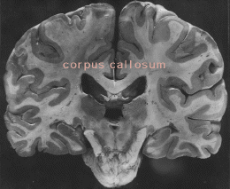
The largest of these is the CORPUS CALLOSUM.
Almost all the commissural connections of the brain are contained in the Corpus Callosum.
It occupies the central area of the space between the hemispheres. A smaller ANTERIOR COMMISSURE serves the decussation of the olfactory tract fibers as well as connections of the temporal areas of the two hemispheres.
The HABENULAR and the POSTERIOR commissures are related to the stalk of
the pineal gland.
- The function of the pineal gland has yet to be definitely determined.
- It is found in all higher vertebrates.
- Its function is partially determined by the effect of light impulses on the retinas.
- It is composed of the Thalamus and Hypothalamus.
- It is rostral to the Brainstem.
- Connecting the Brainstem to the Cerebrum.
- The Diencephalon, as you already know, is hollow, just like the other structures derived from the Neural Tube. The cavity of the Diencephalon is the Third Ventricle.
- The Thalamus serves multiple functions of integration.
- It is best known as the relay station for all incoming sensory impulses except olfaction (smell).
- It appears to have some emotional function and other association functions as well.
- The Thalamus is also a coordinating mechanism for other systems of the CNS. There are relays between the Thalamus and the Cerebral Cortex, as well as between the Thalamus and the Cerebellum and the Thalamus and Basal Ganglia.
- Major ascending (afferent) pathways run through the Thalamus, connecting there to the perceptual centers of the Cortex. Two of these tracts are the Spinothalamic Tract and the Trigeminothalamic Tract.
- The tiny HYPOTHALAMUS lies medial to the ventral portion of the Thalamus. It is a center for the integration of visceral and somatic input, and has connections to that part of the frontal lobe associated with personality. The Hypothalamus influences food and water intake, control of body temperature and certain sexual behaviors. Hypothalamic connections also occur with the nuclei of the Cranial Nerves. These give rise to emotional facial expression.
- These are important in the refinement of movement.
- They appear to accomplish their functions by regulation of gross impulses from other parts of the CNS.
- Perhaps this function is augmented by special synaptic properties.
- Special neurotransmitters.
- Special spatial and temporal interrelations.
- Basal Nuclei synapses have special Neurotransmitters. We will examine neurotransmitters in the section on microanatomy.
- The Basal Ganglia form an important part of the involuntary motor system.
- This is the posterio-medial portion of the Occipital Bone.
- The Brainstem is said to be a "Subtentorial Structure" meaning that it lies below that part of the Dura Mater called the Tentorium Cerebelli.
- The Brainstem is connected to the Cerebellum by three large fiber bundles: the inferior, middle and superior Cerebellar Peduncles.
- The cavity of the brainstem is the Fourth Ventricle. From the Fourth Ventricle, Cerebrospinal Fluid flows out into the Subarachnoid Space.
- There are long ascending pathways which pass from the Spinal Cord through the Brainstem to reach the Thalamus. These include the Spinothalamic Tracts and the Posterior Column-Lemniscal Tracts
- Other pathways descend through the brainstem on their way from the Cerebral Hemispheres to the lower levels. Included among these are the Corticospinal and Corticobulbar Tracts.
- Still other pathways extend from the Basal Nuclei (Ganglia; Striate Bodies) to the Brainstem; to/from the Thalamus to interconnect various CNS regions.
- The major structural components of the Brainstem are the RETICULAR FORMATION the MIDBRAIN, PONS, and MEDULLA (in rostral-to-caudal order).
- The Reticular Formation is the one of the most important features of the Brainstem.
- It is a rather loosely knit group of special neurons is located in the Brainstem throughout its core.
- It consists of multiple neuronal circuits.
- These are arranged in series and in parallel formations.
- The three main anatomical divisions of the Brainstem are the Midbrain, Pons and Medulla.
- Each occupies about 1/3 of the entire structure.
- The Midbrain connects the stem to the Thalamus, and the Medulla links into the Spinal Cord.
- Nuclei The cell bodies of the cranial n.n. are also found in the brainstem.
- These peripheral n.n. serve motor and sensory functions for the head and neck.
- Unlike their spinal nerve counterparts, they may carry sensory or motor or both functions.
- It overlies the brainstem.
- It is connected to the brainstem by three large fiber systems: the Inferior, Middle, and Superior Cerebellar Peduncles.
- Its primary functions involve "UNCONSCIOUS PROPRIOCEPTION."
- Unconscious Proprioception is the nervous system's way of helping the body produce smooth, coordinated and precise movements without our conscious effort.
- The Cerebellum regulates bodily movement by coordinating input from fusal receptors with input from the special senses.
- It receives proprioceptive input from tendons, joints m.m. and semicircular canals.
- It is dominated by the Cerebral Cortex.
- It has efferent and afferent pathways.
- It provides loops that feed neural information back to the origins within the Cerebellar Cortex. Cerebellar connections are complex and include cortical as well as bulbar neurons.
- A Cerebellar hemisphere exerts its control over the ipsilateral extremities.
- It mitigates muscle function through facilitation and inhibition of opposing m.m. groups.
- These groups are comprised of AGONISTS and ANTAGONISTS.
- They oppose each others' actions with SYNERGISTIC function. That means when one group pulls, the other relaxes. Ideally, synergy happens with precision timing, thanks, in part, to the Cerebellum.
- The cerebellum neither initiates movement nor alters sensation but it is essential in...
- Maintenance of postural control.
- Regulation of movements of the extremities
- Regulation of movements of the m.m. speech.
Once you have finished you should:
Go on to Online Lesson 3
or
Go back to Central Nervous System I
E-mail Bill Culbertson
at bill.culbertson@nau.edu
Call Bill Culbertson
at (520) 523-7440
Copyright © 1999
Northern Arizona University
ALL RIGHTS RESERVED