SPH405
| Neurological Foundations of Speech, Language and Hearing |
The Spinal Cord
GOALS:
To present the general form and function of the spinal cord and relate it to
communication.
OBJECTIVES:
Differentiate the gray matter and white mater functions of the spinal cord.
Relate the following spinal cord tracts and nuclei to their functions in human
communication.
- Posterior (dorsal) columns
- Fasciculus Cuneatus
- Fasciculus Gracilis
(Fasciculus Gracilis, Cuneatus)
- Spinothalamic Tracts
- Corticospinal Tracts
The Spinal Cord at the Cervical and Thoracic Levels
The SPINAL CORD is the extension of the CNS outside of the skull.

It consists of an outer layer of myelinated fiber bundles and an inner layer
of unmyelinated neurons.
It is divided from the brain at the level of the FORAMEN MAGNUM.
This is a GEOGRAPHIC (rather than functional) landmark.
This suggests that, anatomically, the spinal cord is just an extension
of the brainstem ( apx. 17inches. to L-1/L-2)
The cord is contained in the vertebral canal of the spinal column.
Open the link below to view how the spinal cord is contained in the spinal column. It will take a few minutes for this movie to download. When it has downloaded, use your mouse to move the spinal cord graphic around to see the spinal cord from different angles.
It is divided into sections that correspond roughly with the vertebral
sections.
At the lower end, spinal segments and vertebral segments diverge.
The infant cord nearly fills the entire length of the vertebral
canal.
As the individual grows, the cord stays the same length, and
the canal gets longer.
So the adult cord extends to the space between L-1 and L-2.
Nerves having their synapses at lower thoracic through
lumbar and sacral segments emerge from the spinal column at
increasingly lower vertebral levels.
Cross sections of the spinal cord reveal a Gray Matter inner portion surrounded by
a white matter outer portion.

The gray matter center of the cord is shaped like the letter H. The "H" extends in three dimensions down the entire length of the
cord. That is, there are two sets of "Horns" and a "Cross Bar." Lets
call the cross bar the "gray commissure." We'll call the horns the posterior or dorsal horns and the anterior or
ventral horns. There are two small lateral horns in the middle, where the
commissure crosses. There is also a white commissure that conveys impulses from one
side of the cord to the other side.
The gray matter of the spinal cord has some mediational function.
This is referred to as the reflexive level of nervous system
function. It is not conscious.
Impulses from the internal organs are generally mediated around the lateral horn and impulses from the voluntary musculature are generally mediated around the ventral and dorsal horns. The white Matter portion consists of the main ascending and descending tracts to and from the higher CNS levels. In general, afferent impulses are conveyed via the dorsal portions of the cord, and efferent functions are conveyed via the ventral portions.
Afferent impulses are visceral, from the internal organs, or somatic,
from the body wall and extremities.
Somatic impulses are exteroceptive or proprioceptive.
- Exteroceptive impulses are stimulated by changes in the outer environment.
- Proprioceptive impulses arise from sensors in the muscles, tendons and joints.
Efferent impulses are also visceral or somatic.
- Visceral impulses are autonomic and move the smooth muscles.
- Somatic motor impulses move the voluntary musculature.
The cord is enlarged at the thoracic and lumbar levels because of the extra mediation needed at the extremities.
The outside white matter of the spinal cord is the main cable for conveying
ascending and descending neural impulses to and from the brain.
- These tracts convey voluntary and involuntary movement.
- Also conscious and unconscious sensations.
- Ascending and Descending Tracts convey action potentials to and from the higher levels, respectively.
ASCENDING TRACTS convey conscious and unconscious sensations.
POSTERIOR COLUMN: Located between the dorsal horns. Impulses to be interpreted as discriminatory tactile, vibratory or position sense.
PINOTHALAMIC: Located in the ventral part of the white
matter cord.
-Conveys Afferent impulses of gross touch, pain and
-Impulses arise in the contralateral side of the body.
DESCENDING TRACTS convey motor impulses to the body and the extremities. Motor impulses: (efferent) cause muscles to contract.
Functions of the Descending Tracts
CORTICOSPINAL TRACTS: Two branches; Lateral and
anterior.
- The Lateral Corticospinal tract in located in the
postero-lateral white area of the cord. It conveys
motor impulses to the extremities.
- The Anterior Corticospinal tract is located between the
ventral horns gray. It conveys motor impulses to the
axial musculature.
There are many other ascending and descending tracts that are
beyond the scope of the present lecture.
- IE: Rubrospinal; Tectospinal; Tegmentospinal; Reticulospinal tracts in lateral columns:
- Relay impulses from higher CNS regions to spinal cord autonomic functions.
- Vestibulospinal tracts: anterolateral; anteromedial in cord.
- These tracts are concerned with balance and "righting" reflex.
Roles of spinal cord in communication:
In Respiration for speech.
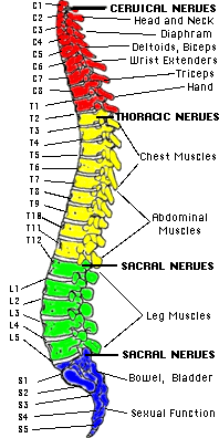
- Cell bodies for the PHRENIC NERVE are located at segments C - 3 4 & 5.
- Tracts for the other m.m. respiration, like the intercostals, pass through this level.
Just for fun: The Phrenic Nerve and Hiccups
In movements of the upper extremities: Thoracic Spinal Cord and brachial
plexus.
- For writing
- For gesturing
Nerve Groups of the Spinal Column Click on "Cervical Nerves."
In movements of lower extremities: Lumbar and sacral levels apply to lower trunk
and extremities.
- These levels have application to accessory m.m. of respiration.
- Perhaps some ancillary aspect of communication.
Once you have finished you should:
Go on to Web Activity 1
or
Go back to Central Nervous System Part I
E-mail Bill Culbertson
at bill.culbertson@nau.edu
Call Bill Culbertson
at (520) 523-7440
Copyright © 1999
Northern Arizona University
ALL RIGHTS RESERVED